Showing posts with label canine anatomy. Show all posts

7/15/12

Segmenting the Canine skull, part 2

I feel like I got a goodly amount of work done on the skull today.  Even if I'm still floundering a bit on where suture lines are. 

Canine skull, end of day 2

I have around 32 different photo references I've pulled from the net, including one where the different bones of the skull have been painted and labeled. 

A very useful powerpoint from the Calgary Association of Veterinary Students.  This is a screenshot from a slide.  They also have the same skull with dorsal, ventral, and rostral views.  I refer to this a lot.


Of course, every skull has some amount of variation.  And I'm not even sure of the breed of the dog scanned.  However, it is a long snout breed, at least.  Those seem to be more common on the internet as well.  I have the feeling that the dog I have is an older example, as sutures tend to become less distinct as individuals age.

I have succeeded in pulling out the mandible, mandibular teeth, nasal bone, incisive bone, zygomatic bone, maxillary teeth and most of the maxillary bone so far. Truthfully, I've been having a hard time even getting a comprehensive list of all of the bones that make up the skull.  It's somewhere between 22 and 50...  Ah, google.

So, I have done three things to rectify my lack of knowledge.  First, a library!  I found a library on Stanford campus that has numerous canine vet books that I should be able to check out.  Second, Amazon! I also found a fairly inexpensive book (creatively called Dog Anatomy, by Peter Goody) on amazon that I decided to buy... I must admit that I turned to Amazon before the library, so technically these are in the wrong order.  However, the prices on Amazon made me sit back and realize that there are much better ways I would like to spend my money, bibliophile tendencies aside.  Which leads me to my third choice, eBay!  Yes, I am now the proud owner of a dog skeleton.  This makes me happier than I probably should be.

My text reference should arrive on Tuesday.  And in the mean time, I've been segmenting. 

Putting the roots on the mandibular teeth.
The newly added mandible and mandibular teeth.
Trying to figure out the nasal and incisive bones.  I can't tell you how tricky those were.

Adding the maxillary bone.  My skull is starting to look colorful!

This is often how I work - multiple views, with the 3D preview to the side.  Because I am having such a difficult time spotting the suture lines, I am relying upon a combination of CT and 3D to figure out where different bones are.  This is why I feel an actual skull is so necessary, at least for me.  Then I will be able to trace the sutures on the physical specimen.

I'm off to the library tomorrow, to further my knowledge of canine anatomy.  I do feel a bit nervous, having started this process without hearing from the actual official people... And truthfully I got a little segmentation happy.  I meant to write my proposal this weekend, not segment canine skulls.  So, realistically I should take a step back and figure that out.  Ick.


7/14/12

Segmenting the Canine Skull

Despite not actually heard back from anyone official on the project idea, I decided to do some segmenting.  Yes, this is what I do for fun on a Saturday night.

The bane of my segmenting existence - ethmoid type bones, and turbinates
I am not looking forward to the canine nasal cavity.  There are a lot of delicate structures in there, that are very, very hard to segment out in Amira.

Canine skull, from an awesome person on deviant art
I have started on other, potentially easily to identify bones on the skull.  However, I've already run into issues there.  The scan I am using, while beautiful, doesn't have the clearest suture lines.  I'm switching back and forth between the three views (sagittal, coronal, and axial) and often viewing the scan side by side with the projected 3D model.  This allows me to see my changes in real time, so I can compare with reference texts and photos.


Amira 3D point cloud

Trying to identify the jugal bone
And the bones of the canine skull are different than those of a human.  So, while the experience I gained segmenting out many, many skulls at my previous job is helpful here, I am struggling somewhat with where suture lines are supposed to be.  I would love to get my hands on an actual skull.  Which is an idea to run by my vet expert....

EDIT: 4 hours later, I'm starting to feel some concern.  I chose what I thought would be 'easy' bones to start off with, but... it has been around 3-4 hours of work and I'm just now finishing them up.  And I'm fairly certain that the lacrimal bone is in there somewhere.  I'm hoping that I just misjudged what would be less difficult, and not that the rest of the skull will be even more complex.  I definitely need a physical canine skull.  Photo references are all well and good, but an actual skull would be much more useful.

Zygomatic bones in red.  (It would be helpful if all of my references agreed on the bone names.) 


7/13/12

Graduation?

One of the things that I regret the most about my time at UIC: BVIS - not finishing.  I went through the (awesome) program, but I never technically finished.  I left for the great California wilds with the research project undone, and it has truthfully been a source of shame for me.

One of those times when you speak up proudly, but mumble the last bit: "Yes, I went to UIC, *but... mumblemumbleresearchprojectunfinshedmumble*."

So!  I am going to finish.  And to aid in that very doable endeavor, I'm going to blog about it as well.  I know it's going to make for scintillating reading.

To start off, I emailed John, the director of the program at UIC now, to find out if I could still graduate at all.  I figured the worst he could say was 'no.'  But, I recieved both a positive answer and the timeline I needed to work in.  That resolved, I decided to scrap my old research on the "Virtual Heart."


I didn't finish the first time, and I had the feeling that if I tried to continue down that road... it was unlikely to happen this time either.  I need a project that I can really get behind.  One that I am likely to enjoy working on most of the time, to get me through that whole 'have to write a ginormous paper' part.

Not to mention, I would truly have to do the literature over again, as most of the articles I had read at the time are already obsolete.  There have been great strides in the virtual world - personally, I've already learned so much about it!  So, not such a great 'research project' now.

One of the things that John asked me was what would I be researching.  This, surprisingly enough, took me aback.  What would I be researching?  Well.... hmph.  What would I be researching?  What would be novel about my project?  What qualified my idea as research potential?

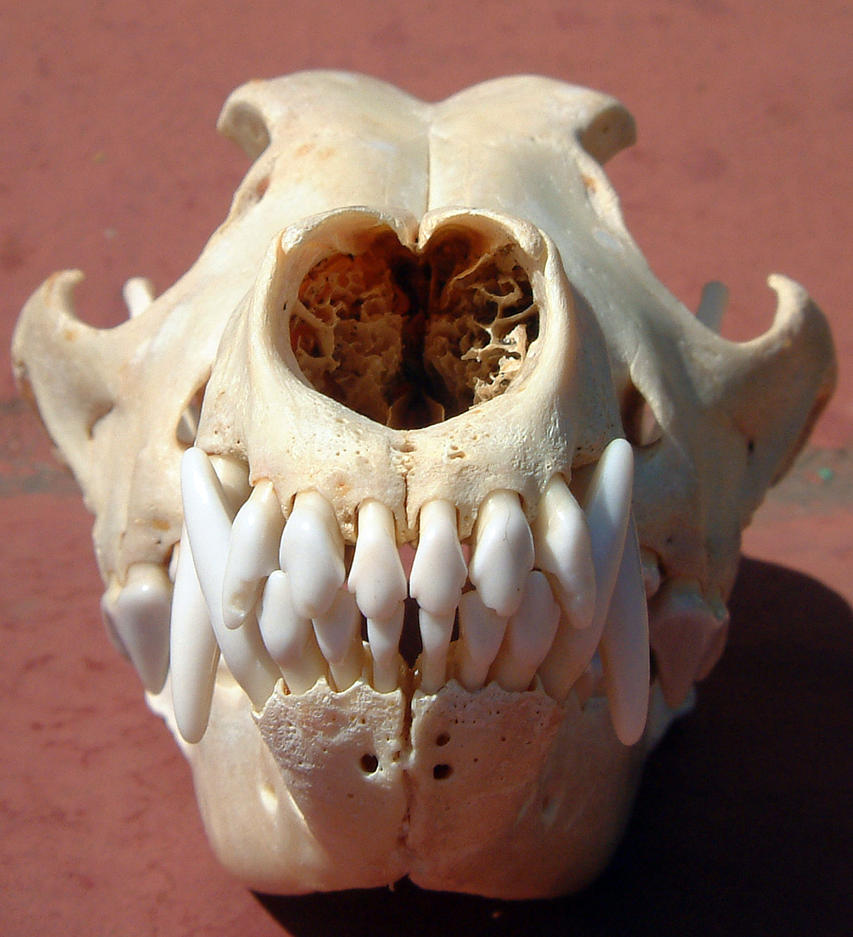
I had this vague idea that I wanted to do something with canines.  And I wanted to concentrate on the skull.  At least most likely.  I had this nebulous concept of 2D to 3D to real time... but then, what would I be researching?  I needed a second opinion.  And I needed to find out what the situation in the veterinarian world actually was.

This was starting to sound like the beginning of a literature review...

I started to research, and it looked very likely that a new type of education was very much needed in the vet world.  But I really wanted to hear from someone close to a 'source.'  So, I was able to get in touch with a Dr. Spriet, from UC Davis, after a bit of perseverance.  I learned that just because people can get very busy, that doesn't mean they are personally ignoring you.  Don't get discouraged.  We had a great talk about the clinical relevance of different projects, the state of veterinary education now, etc.  And I have a much firmer idea of what type of project I want to do now.

So, now I 'just' need to get cracking on a better proposal and then start my literature review.  Have I mentioned that I dislike writing papers? 

The short proposal of a proposal:

Veterinarian programs have been seeing a significant decrease in the number of hours devoted to animal cadaver dissections, turning instead to DICOM slices, and potentially incorporating pro-sections.  This further complicates the learning of complex areas of anatomy, such as the cranial nerves.  Understanding the cranial nerves and their location is especially important when diagnosing node disease in canines, and has been identified as a key difficult study point.  Currently the anatomy of the cranial nerves in canines is taught using a combination of CT and MR slices with basic annotations, power points, journal papers, and anatomy text books.  Cranial nerves are difficult to identify on CT due to contrast resolution, but the bone window for the skull foramina can be located.  Comparing that with the MR slices allows cranial nerves to be located in the most basic sense of the word.  And with actual dissection of the animals becoming much more rare due to time and money constraints, students find the spatial awareness of where cranial nerves are to be hard to understand.

I propose to use a combination of high resolution CT scans and MRI data to build an interactive 3D model of the bones of a canine skull and it's cranial nerves.  The individual bones would be identifiable, as well as the major landmarks and foramina.  The cranial nerves would be added in using Maya, and also be interactive and identifiable.  The structures can be labeled and annotated to increase student knowledge.  Pertinent labels will be identified using the expert help of Dr. Spriet, from UC Davis.  A slider with the CT and/or MRI slice data (superimposed over the 3D model for comparison) will be included to facilitate learning the spacial cues in both 3D and 2D.

Cranial nerves have not been well identified for veterinary students, especially not in an interactive 3D environment.  Combining the DICOM data with the 3D models is a very interesting area of study, and not well researched at this point.  Pairing the two would be a great step forward in the 3D anatomy world as a whole.  While a few people have been able to achieve the combination of a volume rendering of a CT dataset and a 3D surface model, I have not seen slice data combined with a 3D surface model.  However, this is the more likely way a vet will be viewing the data in the practice.  So, combining these learning tools would be of a great benefit to veterinary students.  The process of this development would be CT/MR segmentation (Amira) to 3D (Maya/3D Coat) to Realtime Interactive app (Unity3D).

Obviously, I still have areas to research for my literature review.  I need to better understand canine cranial nerve anatomy, document 3D as a learning tool, and start to research how to begin to combine DICOM with 3D surface models.

Dr. Spriet was also interested in comparing equine cranial nerves with canine, but I was worried that taking it to that level may be overreaching on this project.  I think I could do the segmentation, but I have the feeling that researching and coding the combination of CT and surface models will be an interesting undertaking.  I may be completely wrong, but that would be my initial assessment.

6/29/12

Dog Anatomy 3

So, I fall afoul of deadline dates once again.  I had to submit my work on the canine Anatomicae contest this morning - and I don't feel like I've finished my final piece.  However, isn't the cliche "when do artists actually feel like they've finished?"  Truthfully, there have been some pieces that I really do feel have been completed.  But, this is no where near one of them.

Some WIP shots.

Color blocking in the silhouette and choosing some colors to work from

Basic shading added

adding in a very rough background and refining the shading - her paw looks amputated right now

tried to work on her eyes and the paws.  Got some reflected light going on, but it's not quite there.

The image itself is still very rough - blocky shading and such.  Her eyes aren't quite right, and the background is atrocious.  But, this is where I got to in two weeks time, while also working and doing the paper mache eye, etc. 

The contest itself required a pose that showed all four legs.  I wasn't completely aware of that when I did my anatomy sketches... But I do plan on including the anatomy for this pose as well, when I get the time.  Perhaps in between paper mache layers?

- SH